INTRODUCTION
Hamstring muscle strains are often observed in sprinting and jumping activities (Woods et al., 2004). The incidence of hamstring strain is 12%–16% of all injuries and 54.4% of all muscle strains in athletes (Eirale et al., 2013; Ekstrand et al., 2011). Risk factors of hamstring strains include: previous hamstring injury, improper warm up, core muscles weakness, fatigue, low coordination of trunk and pelvic muscles strength, lumbar posture abnormality and imbalance between muscles strength ratio (Cameron et al., 2009; Schmitt et al., 2012; Sherry and Best, 2004; Yeung et al., 2009).
Hamstring injuries often occur with extreme stretch in simultaneous hip flexion and knee extension during eccentric contractions (Askling et al., 2007). Also, rapid change of hamstring contraction from eccentric to concentric can cause hamstring injuries (Woods et al., 2004). Eighty percent hamstring strains occur in long head of biceps femoris (BF) during terminal swing, hence conception of causes and hamstring injury prevention is important for athletic society (Chumanov et al., 2007; Koulouris and Connell, 2003).
Long head of BF and gluteus maximus (Gmax) stabilize pelvis (Leinonen et al., 2000) and Gmax keeps upright position in standing (Jenkins and Hollinshead, 1998). Similarly, erector spine (ES) muscles keep trunk erect posture during high speed running (Sado et al., 2016) and internal oblique (IO) muscles activity decreases stretch on BF (Devlin, 2000). Sagittal plane movements of pelvis and hip in subjects with previous hamstring injury are asymmetry (Daly et al., 2016). Furthermore, the synergist muscle weakness on the same joint increases activity demands compared to other synergists, then muscle strain may occur (Sahrmann, 2002). Gmax and ES activity as trunk extensor synergists changes Gmax to ES activity ratio (Gmax/ES ratio) (Kim and Yoo, 2016). Also, the decreased Gmax activity relative to semitendinosus (ST) muscle was related to increased ipsilateral ES activity (Tateuchi et al., 2012).
The nordic hamstring exercise (NHE) is a progressive eccentric exercise that is simulated a condition with high muscle force demand during knee extension (Iga et al., 2012). This task reduced the incidence rate of primary hamstring injuries to 60% (Thorborg, 2012). Hamstring activation on two joints during NHE increases effectively maximal eccentric hamstring strength more than traditional exercises (Mjølsnes et al., 2004). The chronicity of agonist/antagonist imbalances has the important role in occurrence of recurrent hamstring injuries (Croisier et al., 2002; Croisier, 2004). In previous studies the preventive and treatment methods have been examined only in chronic stage of hamstring strains (Croisier et al., 2002). Hence, rehabilitation exercise programs must be individually planned for preventing of recurrence chronic injuries (Croisier, 2004).
The impairment of abdominal, Gmax and rectus femoris (RF) muscles action can make overuse syndrome of hamstring muscles, too (Sahrmann, 2002). Thus, it is very helpful for injury prevention to find how other muscles on hip and knee joints act (Sahrmann, 2002). Change in knee joint angle from flexion to extension increases abdominal muscles activity to control of trunk (Eom et al., 2016). Furthermore, antagonistic cocontraction of trunk muscles provides mechanical stability of lumbar spine in neutral postures (Cholewicki et al., 1997). Therefore the increased spinal loads increase abdominal and back muscle cocontractions (Granata and Marras, 2000). Hip and core stability provides a stable base for lower extremities movements (Willson et al., 2005). Likewise, proper function of anterior and posterior trunk and hip muscles in sagittal plane is necessary for core stability (Willson et al., 2005). Therefore, core dysfunction increases the risk of lower extremity injuries (Willson et al., 2005).
In previous investigations have not been shown trunk and hip muscles activity levels during hamstring contractions and there was a lack of study about the role of hip and trunk muscles during NHE. Also, some studies were concentrated on hamstring/quadriceps muscle activity ratio (H/Q ratio) but there wasn’t any study about Gmax/ES ratio during NHE.
The main purpose of this study was to analyze of hip and trunk muscles activity during NHE. We hypothesized that trunk muscles activity level would increase and Gmax/ES ratio would decrease with increasing peak knee extension angle during NHE.
MATERIALS AND METHODS
Subjects
Ten healthy men (age, 26.1±5.46 years; height, 172.9±6.57 cm; weight, 60.7±5.79 kg; body mass index, 20.32±1.53 kg/m2); participated in this study. If the subjects had neuromuscular, cardiovascular disease and hamstring injury during the previous 6 months, they were excluded from this study. Ethical approval was obtained from Waseda University.
Experimental protocol
Prior to measurements, the subjects were instructed how to accomplish the NHE. The starting position of NHE was kneeling with upright state of trunk over a mat on the ground, knees flexed 90° and both arms placed in front of the body. A cooperator was holding lower legs of the subject to support. The participants lowered whole of trunk toward the ground as far as possible without bending or rotating in hip joint or trunk while keeping neutral posture in trunk and hip joints. The participants were allowed to incline until maximum downward moving point. When the participants reached the maximum point they returned back to the starting position without changing in straight status of the body. They performed this NHE movement two repetitions in a row with 2-min resting time between trails and two sets of the task. A metronome was set at 60 beats per minute to define the movement speed. Participants leaned forward as much as they could for 3 sec and came back to start position for another 3 sec. Participants performed NHE twice in a row then 2-min resting time was allocated between trials. Manual resistance was applied for each muscle to obtain maximal voluntary isometric contractions (MVICs) for 3 sec after NHE trials.
Electromyography and kinematics measurements
We examined electromyography (EMG) characteristics of fifteen muscles. The muscles in right (Rt) side of body were included: BF, ST, Gmax, gluteus medius (Gmed), RF and in Rt and left (Lt) sides of body: IO, external oblique (EO), rectus abdominis (RA), multifidus (MF), and ES. After shaving and cleaning the skin with alcohol, we used bipolar surface Ag/Agcl electrodes (Blue sensor N-00-S, METS Co., Tokyo, Japan). The diameter of surface electrodes was 8 mm and interelectrode distance was 20 mm. A wireless EMG telemeter system (Biolog DL-5000, S & ME Co., Tokyo, Japan) were used to examine muscle activities.
Location of muscles was confirmed with palpation of the landmarks and isometric muscle contractions. The electrodes placement on both side of spine was for ES; 2 cm lateral of the 3rd lumbar spinous process and MF; at level of 5th lumbar spinous process on a line extending from posterior superior iliac spine to first lumbar vertebra. The electrode placement was for Gmax; the midpoint between greater trochanter and sacrum and Gmed; the midpoint between iliac crest and greater trochanter of femur. Also, the electrodes were placed for ST; the midpoint between ischial tuberosity and medial epicondyle of tibia, RF; the midpoint between anterior superior iliac spine and superior edge of patella, and BF; the midpoint between ischial tuberosity and lateral epicondyle of tibia. For abdominal muscles, along the fibers electrodes placement were for IO; 2 cm medial side of anterior superior iliac spine, EO; 15 cm lateral side of umbilicus and RA; 3 cm lateral side of umbilicus (Hermens et al., 2000).
Kinematic data were recorded using 8 motion capture cameras (Oqus, Qualysis Ltd., Co., Goteborg, Swedish). The reflective markers were placed in Rt side of body on greater trochanter, lateral condyle of femur and lateral malleolus. The knee joint angle was defined between the lateral condyle of femur and along of thigh and shank. The knee joint angle was calculated as motion of the thigh relative to shank using the reflective markers of motion capture cameras. Also, peak knee angle for each subject was calculated by subtracting the angle between thigh and shank where the subject could incline maximally downward (the peak knee extension angle) from the 90° angle at starting position.
Data analysis
The sampling rate of EMG was 1,000 Hz and it was 200 Hz for motion system. Raw EMG signals were analyzed with software (BIMUTAS-Video, Kissei Comtec Co., Ltd., Nagano, Japan). The EMG data were rectified, high-pass filtered at 20 Hz, and low-pass filtered at 500 Hz to remove the artifacts. The 2-dimensional angles of knee in sagittal plane and muscles activity were the dependent variables. A trigger mechanism was used to synchronize the EMG measurements and the motion capture system data.
In this study, NHE was included downward and upward motions. Downward motion was defined as forward movement from starting position or kneeling toward ground until maximum knee extension angle that each subject could achieve with knee extending. Upward motion was defined as backward movement while knee flexing from maximum knee extension angle toward starting position. The muscles activity levels were measured in 1 sec before maximum knee extension angle during downward motion and in 1 sec after maximum knee extension angle during upward motion. Therefore the onset of motion in downward motion was from kneeling with the point defined between the markers on greater trochanter, lateral femoral condyle and lateral malleolus and the end of motion was where the participants could move maximally downward (peak knee angle). Also in upward motion, the onset of motion was reversely from the peak knee angle and the end of motion was kneeling. The root mean square (RMS) values of each muscle over 1 sec in downward and upward motions were normalized by RMS values of MVICs in each muscle to calculate %MVIC. The EMG signals were designated less than 10% MVICs to reduce probable signal changes with fatigue and the electrode displacement.
Statistical analysis
The data were analyzed in SPSS ver. 17.0 (SPSS Inc., Chicago, IL, USA). We used the parametric statistical methods because all data had a normal distribution confirmed by using Kolmogorov–Smirnov test. We analyzed muscles activity using repeated measure analysis of variance to compare muscle activity levels in downward and upward motions, separately. Also, we compared muscle activity levels with paired t-test between downward and upward motions. Statistical significance was determined as P<0.05 for all statistical tests. We used Pearson correlation to determine relation between Gmax/Es activity ratios and peak knee extension angle. We calculated Gmax/ES ratio in downward and upward motions. Also, we examined the correlation between Gmax/ES ratio and peak knee extension angle in both motions during NHE.
RESULTS
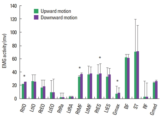
The results of Mean and standard deviation of muscles activity levels in downward and upward motions during NHE are presented in (Table 1, Fig. 1). Mean EMG activity level of ST muscle in downward and upward motions during NHE was the greatest of all muscles and BF activity level was the second greatest (P< 0.05) (Table 1). Considerably, ES and MF muscles activity levels were greater than other muscles after hamstring muscle activity. Also, IO and EO muscles activity levels were greater than other trunk muscles after ES and MF muscles, respectively (P<0.05).
There were significant differences in RtIO, RtGmax, RtMF, and ES muscle activity levels between downward and upward motions during NHE (P<0.05) (Fig. 1). Therefore, RtIO, RtGmax, RtMF, and ES muscle activity levels during upward motions were significantly greater than those in downward motion. The mean difference of RtMF activity level between two motions was greater than other muscles activity levels (P<0.05). Mean peak knee angle was 72.87±6.70 for all subjects during NHE.
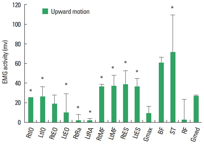
The EMG muscles activity levels in downward motion during NHE are shown in (Fig. 2). RtIO and LtIO muscle activity levels were more than RtRA, LtRA, and RtRF muscle activity levels (P<0.05). Also RtMF, LtMF, RtES, and LtES muscle activity levels were more than LtEO, RtRA, and LRA muscle activities (P<0.05). MF and ES muscle activity levels in both sides were more than RtGmax and RtRF muscle activities (P<0.05). LtIO muscle activity level was more than RtRF muscle activity (P<0.05) (Fig. 2).
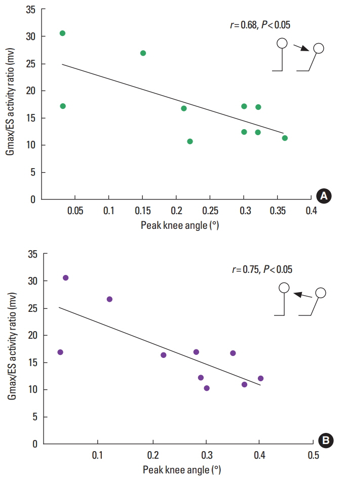
Also, the EMG muscle activity levels in upward motion are shown in (Fig. 3). RtIO and LtIO muscle activity levels were more than RtRA, LtRA, RtRF, and RtIO muscle activity level was more than RtGmax in upward motion (P<0.05). RtMF and LtMF and ES muscle activity levels were more than LtEO, RtRA, and LtRA muscle activity levels (P<0.05). Therefore, IO, ES, and MF muscle activity levels were significantly increased after hamstring activity in both upward and downward motions (P<0.05) (Figs. 2, 3). Gmax/ES activity ratio was 0.22±0.11 in downward motion and 0.24±0.13 in upward motion. A significant negative linear correlation was found between Gmax/ES ratio and peak knee angle in downward (r=0.687, P=0.028) and upward motions (r=0.753, P=0.012), respectively (Fig. 4).
DISCUSSION
This study is the first analysis of low back, abdominal, hip, and hamstring muscles activity in both downward and upward motions during NHE. The highlight finding of this study was that ST muscle activity level was the highest among the muscles during downward and upward motions. Also, trunk muscles activity level especially back extensor (ES) and oblique muscles (IO) was increased and Gmax/ES ratio decreased with increasing knee extension angle during NHE.
The NHE improves eccentric hamstring muscle strength (Delahunt et al., 2016). Eccentric contractions have been used clinically for hamstring strain prevention in athletes (Murphy et al., 2012; Orchard et al., 2013). Although several studies have examined hamstring muscle activity, the assessment of hip and trunk muscles activity was necessary during NHE. We concur the researchers that ST muscle is preferably activated during NHE (Bourne et al., 2016) and confirm the results of (Ditroilo et al., 2013) that BF activity during NHE was very high.
The kinematic and neuromuscular analyses can achieve beneficial issues for coaches and athletes of sport medicine in hamstring injury prevention (Brukner, 2015). Other studies estimated that BF muscle was activated more than other hamstrings in eccentric contraction (Higashihara et al., 2010; Woodley and Mercer, 2005), but in our study ST muscle activity level was the highest during NHE. According to our results, MF and ES muscles activity level after hamstring activity level were more than other muscles in downward and upward motions. Trunk muscles attached to pelvis and control the pelvis tilts and length changes of hamstring muscle. These muscles provide the proper condition for optimal hamstring contraction and prevent injury in high speed sports (Jull and Richardson, 1994; Sherry and Best, 2004; Wohlfahrt et al., 1993).
Isometric ES muscle contraction is necessary for optimal standing position (Boucher et al., 2013). Moreover abdominal and hip extensor muscles with posterior pelvis tilting and back extensor and hip flexors with anterior tilting contribute to force couples for spinal stability (Sahrmann, 2002). Thus coactivation of force couples is an important factor in maintaining neutral pelvis tilt and lumbar lordosis (Granata and Marras, 2000; Jull and Richardson, 1994). Consequently, posterior hip and trunk muscles with central of gravity displacement activate in NHE and these muscles with isometric contractions as antigravity muscles are responsible for keeping erect posture.
Furthermore, isometric abdominal contractions keep erect position of spine during NHE. Inferomedial fibers of IO muscles with force closure mechanism contribute to sacroiliac joint control (Snijders et al., 1995). Also, IO muscles neutralize the pelvis tilts that gravity force creates (Snijders et al., 1998). Regarding previous studies, IO muscle activates greater than EO against gravity in trunk erect position (Snijders et al., 1995; Vleeming et al., 1997) and IO muscle keeps pelvis stability during lower limb movement (Floyd and Silver, 1950). Consequently, trunk muscles coactivate to keep Rt position of spine during eccentric and concentric contractions of hamstrings (Sahrmann, 2002). The researchers showed that imbalances of core muscles caused pelvis instability as hamstring strains will occur during eccentric contractions (Chumanov et al., 2007). Vleeming et al. (1997) and Snijders et al. (1995) showed that oblique abdominal muscles in trunk erect position have greater activity level against gravity relative to Gmax, ES and BF.
In our results, IO muscles activity levels after hamstring and back extensor muscles activity were more than other muscles activity in both downward and upward motions during NHE. Thus, MF, ES and oblique abdominal muscles create sufficient stability in trunk and pelvis and these muscles cause a proper performance basis for hamstring muscles during NHE. In other words, when MF, ES and abdominal muscles activate greater, reciprocally, eccentric and concentric hamstring contractions will be better. The results of this study highlighted the role of IO and ES muscles during hamstring eccentric and concentric contractions. Sherry and Best (2004) presented that trunk stabilization exercises can prevent hamstring strains in athletes. Therefore we conclude that IO, ES and MF strengthening can affect hamstring contractions improvement. These results are very important in design of injury preventive protocols for athletes with more risk of hamstring injury, especially soccer players and runners (Murphy et al., 2012).
Furthermore, we found that Gmax/ES ratio had a negative correlation with knee extension angle during NHE. Gmax activity level decreased significantly with increasing knee extension angle relative to ES activity. In other words, whatever knee extension angle increased, the demand of ES activity was more than Gmax. Guilhem et al. (2014) showed linear correlations between the EMG activity (RMS) and isometric peak torque of ES muscles. Also Sado et al. (2016) showed that hip extension torque and posterior pelvis tilt increase during sprinting in stance limb. ES muscles activate to keep trunk erect posture and neutralize the posterior pelvis tilt and lumbar flexion with increasing lumbosacral extension torque during high speed running (Sado et al., 2016). When the participants inclined downward in NHE, ES muscles activated to counteract the hip extension torque. Moreover, regarding to decrease the distance of ES to ground, ES activated to keep trunk erect posture versus gravitational force and hip extension torque more than Gmax. Similarly, Gmax/ES ratio increased with decreasing knee extension angle in upward motion. In other words, the demand of ES activity with decreasing knee extension and returning to kneeling decreased.
In summary, these results emphasized that the high activity level of ES muscles during eccentric and concentric hamstring contractions is very necessary. However, sufficient contractions of both ES and Gmax are important for pelvis stabilization and proper hamstring performance. Thus, the assessment of Gmax/ES ratio during NHE and strengthening of ES and Gmax muscles can be very important in rehabilitation of muscle imbalances and hamstring injury preventive methods. Also, if each Gmax or ES muscle activate inefficiently, the hamstrings activity may increase compensatory. This explains the synergistic cooperation between Gmax, ES and hamstring muscles in closed kinematic chain of lower limb as other studies (Leinonen et al., 2000; Nelson-Wong et al., 2012). Unlike another studies, H/Q ratio wasn’t decreased in our results, because all our participants were healthy. Also, we did not found other significant muscle ratios in this study. We recommend more studies about muscle interactions and activity ratios.
There were some limitations to this study. We did not examine the effect of movement velocity and the effect of velocity on muscles activity in this study. Some previous studies were demonstrated that the extent of peak hip and knee angles in different velocities was various (Chumanov et al., 2011; Kuitunen et al., 2002; Thelen et al., 2005). Also they concluded that abdominal oblique muscles activity causes to reduce hamstring stretches in different velocities, therefore we suggest that further studies are necessary (Chumanov et al., 2011; Kuitunen et al., 2002; Thelen et al., 2005).
All subjects in this study were healthy and did not have any hamstring injury. Our results as the first step for assessment of hip and trunk muscles performance during hamstring eccentric and concentric contractions are sufficed only. Hence future researches about comparison of relationship between trunk and hip muscles performance in healthy and injured subjects during NHE would be needed.
In conclusion, neuromuscular analysis of NHE demonstrated that high increased ST and BF activity levels in eccentric and concentric contractions compared to other muscles during NHE. Furthermore, increased activity of ES and oblique muscles (IO) and decreased Gmax/ES ratio was related to increased knee extension angle. We recommend that hamstring injury preventive programs and comprehensive rehabilitation after emphasis on hamstring muscles strengthening will be concentrated on trunk and hip muscles.