Tuberculosis of testis and prostate that mimicked testicular cancer in young male soccer player
Article information
Abstract
Staphylococcus infection was the most common organism found in infection of athletics, and tuberculosis (TB) was rare. Although genitourinary tuberculosis (GUTB) was the most common subtype of extrapulmonary tuberculosis (EPTB) in the past, it was recently reported to account for less than 0.5% of all patients with EPTB and 1.5% of all patients with pulmonary tuberculosis (TB). And, there are few cases reported about concomitant tuberculous infection of testis and prostate. Pubic pain is a common symptom in soccer player and its cause can be difficult to determine. A 25-yr-old male soccer player presented with persistent pubic pain of unknown origin. Incidentally, right testicular mass was detected on physical examination. Computed tomography revealed a multiple enlarged retroperitoneal lymph nodes. Under the clinical diagnosis of a right testicular tumor, right radical inguinal orchiectomy was performed. And prostate biopsy was performed due to elevated serum prostate specific antigen (PSA). Pathologic examination confirmed concomitant TB of testis and prostate.
INTRODUCTION
The number of new tuberculosis (TB) patients in Korea was 32,010 (67.2 in 100,000) in 2002 and 30,687 (64 in 100,000) in 2003. In 2008, the number of new patients with extrapulmonary tuberculosis (EPTB) was 5,813, which accounted for 17.0% of all new TB patients. The number of new genitourinary tuberculosis (GUTB) patients was 123 (0.4%) in 2008 and 146 (0.4%) in 2009, which ranked lowest in EPTB (Lee et al., 2011). The term GUTB was first introduced by Willbolz et al, and it represents tuberculosis that occurs in the kidney, ureter, testis, and epididymis through blood-borne infection (Gow, 1971). Although GUTB was the most common subtype of EPTB in the past, it was recently reported to account for less than 0.5% of all patients with EPTB and 1.5% of all patients with pulmonary TB (Jacob et al., 2008). In case of genital TB, nonspecific clinical presentations and variable radiographic presentations may mimic other pathologic lesions (Wise and Marella, 2003). Male genital TB can present as a testicular mass that is difficult to differentiate from malignancy (Jacob et al., 2008). We report a case of TB developed in young and healthy male soccer player. TB concomitantly arose in the prostate and testis, which mimicked testicular cancer. And, we reviewed the related literatures.
MATERIALS AND METHODS
A 25-yr-old male soccer player visited to urology department because of persistent pubic pain. He took several antibiotics and non-steroidal anti-inflammatory drugs (NSAIDs) for several months in some clinics, diagnosed as chronic pelvic pain syndrome or chronic prostatitis. On physical examination, we found a right testicular enlargement incidentally. The patient complained of little testicular pain, and also no generalized or voiding symptoms. Non-tender enlarged right testis with irregular surface was palpated, which was measured about 6×5×4 cm. The epididymis was hard, non-tender, and adhered to the testis, and the vas deferens was normal on palpation. The soft firm prostate was palpable by digital rectal exam (DRE), without any abnormal findings. Chest radiograph was normal, and laboratory data including complete blood cell count, blood chemistry, testicular tumor markers including α-fetoprotein (AFP), β-human chorionic gonadotropin (β-hCG), lactate dehydrogenase (LDH) were all within normal limits. Urinalysis showed pyuria of 3–5 white blood cells per high power field, but urine culture showed no growth of microorganism. His serum prostate specific antigen (PSA) level and prostate volume were 5.23 ng/mL (TandemR-R PSA immunoradiometric assay) and 26 cc, respectively. There were no other abnormal findings except enlarged prostate and prostatic calcifications in transrectal ultrasonography (TRUS).
RESULTS
In suspect of testicular cancer or TB, urine acid-fast bacilli (AFB) stain and TB-polymerase chain reaction (PCR) test were taken, which were all negative. Scrotal Doppler ultrasound (US) showed multiple intratesticular hypoechoic masses with enlarged ipsilateral epididymis (Fig. 1). Abdomen and pelvis CT scan showed multiple enlarged retroperitoneal and pelvic lymph nodes (aortocaval, left paraortic, both common iliac, both external and internal iliac and presacral lymph node) (Fig. 2A). Under the clinical diagnosis of right testicular cancer, right radical inguinal orchiectomy was performed. The pathology of right testis and epididymis showed granulomatous inflammation with caseous necrosis and histiocytic aggregation (Fig. 3). The AFB stains of the scrotal tissues were all negative, but TB-PCR test was positive. TRUS-guided prostate biopsy was also performed due to high PSA. And the pathology of prostate also showed granulomatous prostatitis (Fig. 4). We finally diagnosed the patient as concomitant GUTB involving testis, epididymis, and prostate.
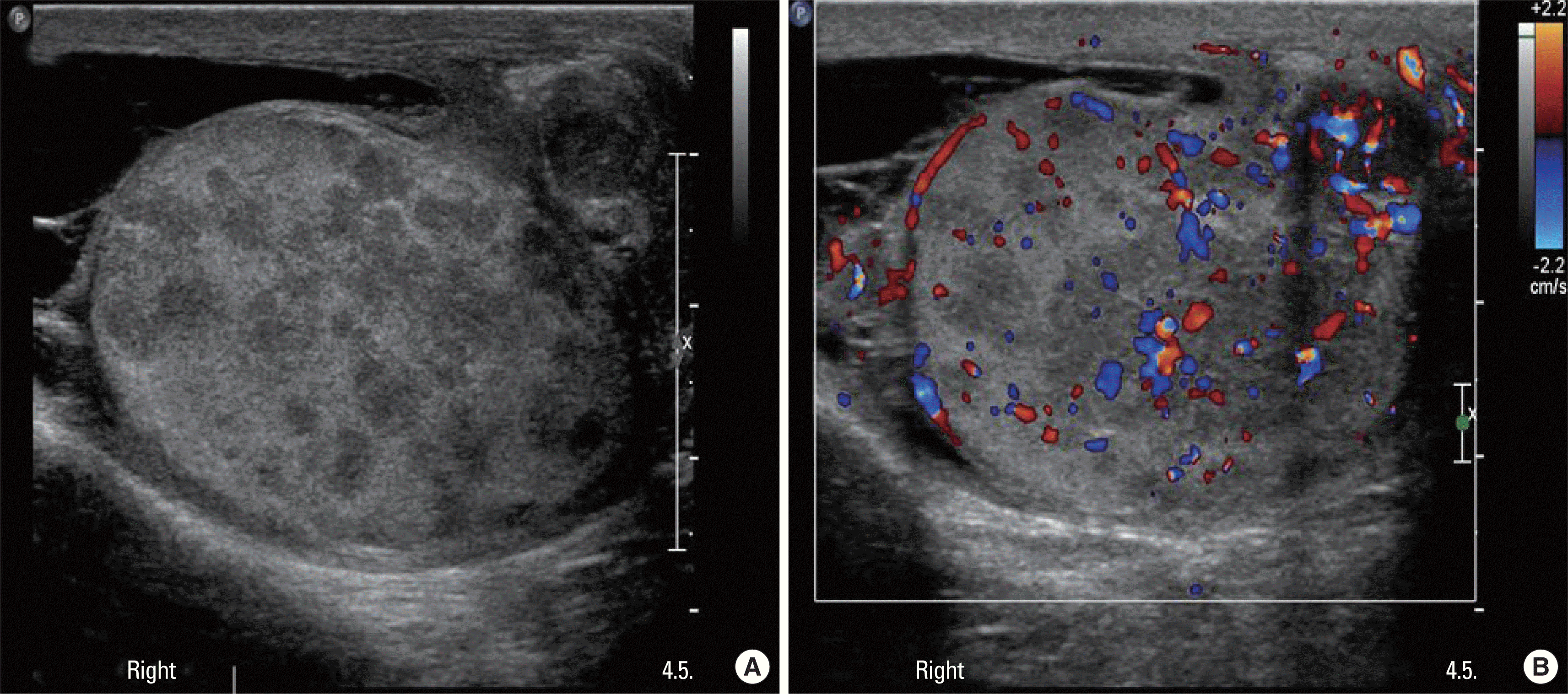
Ultrasonography of testis showing multiple hypoechoic lesions inside the right testis in gray-scale image (A) and color-Doppler image (B).
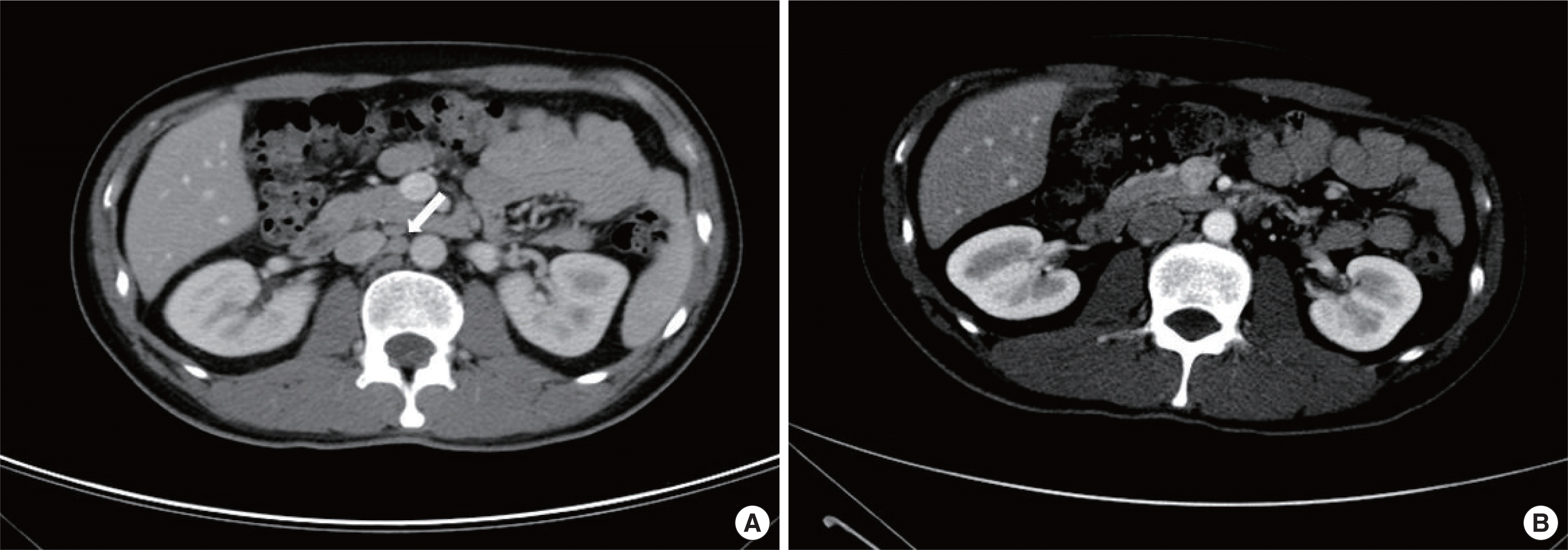
(A) Computed tomography examination showing enlarged aortocaval lymph nodes (arrow). (B) Computed tomography examination after 6 months of anti-TB medication treatment, showing a disappearance of enlarged aortocaval lymph nodes.
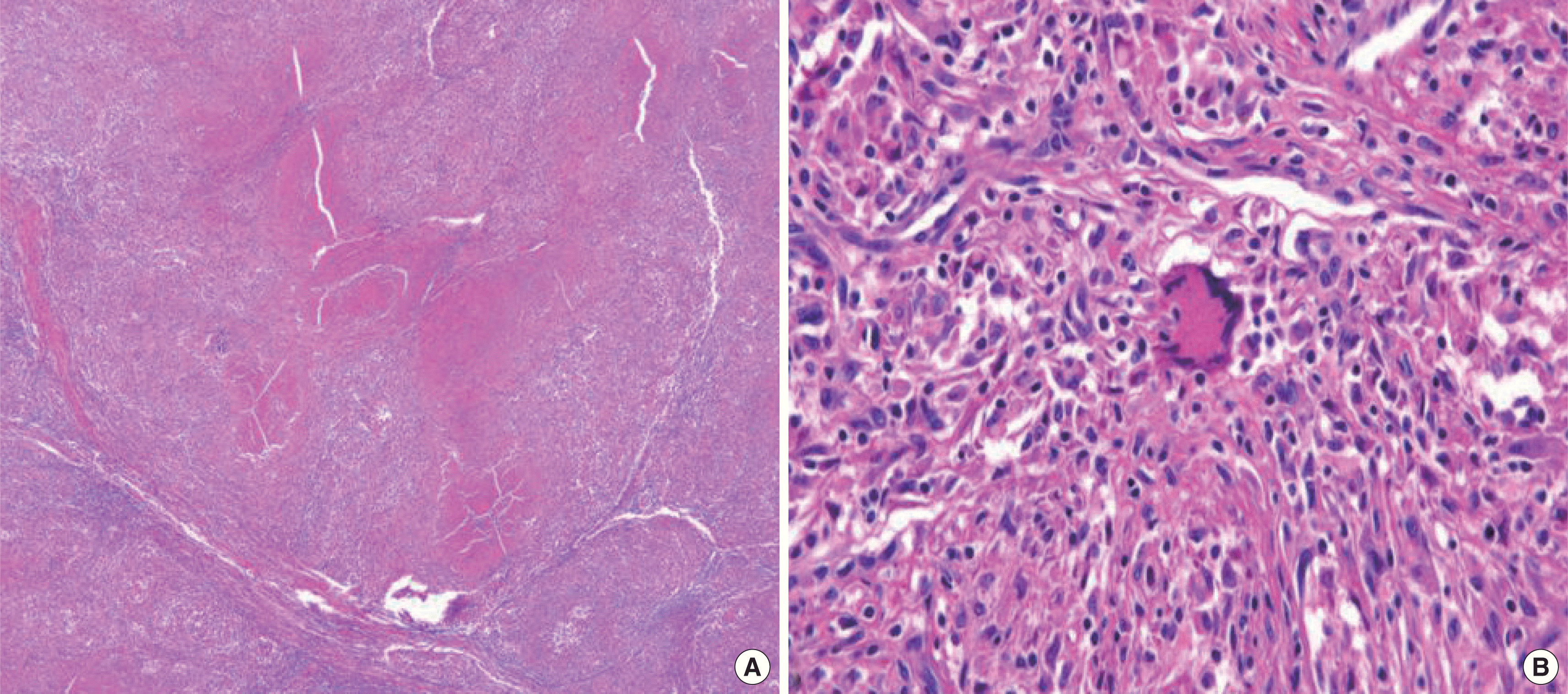
(A) Granulomatous inflammation in the testis, featuring caseous necrosis surrounded by epitheloid cells and lymphocytes (H&E stain, × 100). (B) Microscopic finding show Langhans’ giant cell surrounded by lymphocytes and fibroblast (H&E stain, × 400).

Granulomatous inflammation in the prostate showing central necrosis surrounded by numerous granulomas, some of which contain giant cells in their centers. (A: H&E stain, ×100, B: H&E stain, ×400)
After histologic confirmation of TB, the patient started anti-TB chemotherapy with regimen of isoniazid (INH), rifampicin (RFP), ethambutol (EMB), and pyrazinamide (PZA) for 2 months, following four months of INH, RFP. When finishing the anti-TB chemotherapy, the enlarged retroperitoneal lymph nodes were disappeared on abdomen and pelvis CT scan (Fig. 2B), and serum PSA was normalized to 2.14 ng/mL.
DISCUSSION
Pubic pain is a common symptom in soccer player, but its cause can be difficult to determine and NSAIDS or antibiotics empirically used to improve symptom (Guis-Sabatier et al., 1999).
GUTB recently reported to account for less than 0.5% of all patients with EPTB and 1.5% of all patients with pulmonary TB (Jacob et al., 2008). About 28% of these patients have isolated genital organ involvement (Wolf and McAninch, 1991). Nonspecific clinical presentations and variable radiographic presentations mimic other pathologic lesions (Wise and Marella, 2003). Clinical manifestations are rarely systemic and more likely to localize to the level of disease (Christensen, 1974).
Male genital TB can present as a testicular mass that is difficult to differentiate from malignancy (Jacob et al., 2008). The mode of spread of TB to the scrotum is controversial, but is believed to occur by one of several mechanisms: hematogenous, retrourethral, lymphatic, and direct extension (Wolf and McAninch, 1991). Testicular infections are usually associated with epididymal infections. The disease usually starts in the globus minor which has a greater blood supply than other portions of the epididymis (Kim et al., 2005; Wise and Marella, 2003). TB of the prostate is less common than vesiculo-seminal and epididymal TB (Gupta et al., 2008). The most common mode of involvement is hematogenous, though descending infection and direct intracanalicular extension is also known. Sterile pyuria and hematuria are classic findings of urinary TB (Wise and Marella, 2003). But, most common symptom with prostatic involvement is urinary frequency and nocturia (Sporer and Oppenheimer, 1957). Other urinary symptoms such as dysuria, hematuria, and hematospermia also occur with prostatic involvement (Jacob et al., 2008). On account of its uncommon presentation, many cases are found incidentally following transurethral re-section.
Classically, the diagnosis of genitourinary TB is made by the identification of Mycobacterium tuberculosis in the urine. Urine AFB culture is also used for diagnosis because AFB smears are often negative. Urine AFB cultures, however, take 6 to 8 weeks because M. tuberculosis grows slowly. The use of TB-PCR and nucleic acid amplification has improved sensitivity and specificity in diagnosis of TB. Moussa and colleagues reported the sensitivity and specificity of urine TB-PCR to be 96% and 98% respectively (Moussa et al., 2000).
The treatment of choice is chemotherapy using three to four anti-TB agents for up to six to nine months. Surgical treatment should be considered in cases not responding to chemotherapy or suspected malignancy.
In this case, radical inguinal orchiectomy was performed despite normal testicular tumor markers, because urine AFB stain and TB-PCR were all negative and other clinical manifestations (normal chest radiograph, enlarged retroperitoneal lymph nodes on CT scan, multiple hypoechoic masses on scrotal US) indicates testicular cancer rather than TB. At 6 months after anti-TB chemo-therapy, the enlarged retroperitoneal lymph nodes and serum PSA were normalized. A case of concomitant prostate and testis TB accompanied with multiple retroperitoneal lymphadenopathies is extremely rare. We want to convey the message that in the atypical symptoms and/or signs of patients presenting with testicular swelling and pubic pain, TB should be considered as a possible diagnosis.
Notes
CONFLICT OF INTEREST
No potential conflict of interest relevant to this article was reported.