Characteristics of upper limb muscular strength in male wheelchair tennis players
Article information
Abstract
The purpose of this study was to identify the characteristics of muscular strength in upper limb and to present the preliminary information for development of sports injury prevention program and exercise rehabilitation program in wheelchair tennis players. Participants were 12 male wheelchair tennis players. Muscular strength was measured in shoulder and elbow joints with isokinetic dynamometer. Ipsilateral (IR) and bilateral (BR) balance ratio were calculated with isokinetic strength at 60°/sec. As a result, extension strength (ES) was significantly higher than flexion strength (FS) (P< 0.001), and IR in both sides and BR in ES were maintained within normal range whereas BR in FS was lower than normal range in shoulder joint. In elbow joint FS was significantly higher than ES (P< 0.05), and IR and BR were lower than normal range. Consequently, the different tendency in IR between shoulder and elbow joints and lower IR and BR in elbow joints could be the characteristics in male wheelchair tennis players. It is suggested that flexor strengthening program in nondominant shoulder joint, extensor strengthening program in both elbow joint, and flexor strengthening program in non-dominant elbow joint should be introduced for male wheelchair tennis players.
INTRODUCTION
A wheelchair is a very important transportation method for wheelchair dependents (Curtis et al., 1999). A propulsion cycle of wheelchair is comprised of a pushing stage, where hands are touching handrims and providing propulsion to wheels, and a recovery stage, where they come back to the starting position (Chow et al., 2001). The shoulder joint flexion and the shoulder girdle protraction are shown during the pushing stage, and the shoulder joint extension, the abduction, and the shoulder girdle retraction are shown during the recovery stage in upper limbs (Morrow et al., 2009).
Also, in the first 1/3 section of the pushing stage, middle deltoid is utilized for the first time, and pectoralis major, coracobrachialis, anterior deltoid, and infraspinatus muscles are utilized next. Brachial biceps are mainly utilized for the next 1/3 section, and brachial biceps, brachioradialis muscle, brachialis muscle, pronator teres, and pronator quadratus muscle are utilized in the final 1/3 section (Rankin et al., 2011). For wheelchair dependents, the strength of propulsion of wheelchair itself was reported at or below 50% of heart rate reserve (HRR) except for the uphill propulsion of wheelchair (Coutts, 1988), thus the intensity of comfortable pushing of wheelchair in everyday activities are not enough to improve cardiopulmonary function of wheelchair dependents (Chung and Shin, 2001). But long term, continuous wheelchair propulsion causes scapula protraction, causing relative internal rotation of humerus and elevation due to the repetitive exercise pattern of shoulder joint, elbow joint, and carpocarpal joint (Schantz et al., 1999).
Also, the elevation in the state of adduction and internal rotation of humerus causes the burden in the direction of the long axis of humerus and leads to degenerative changes and soft tissue disorders of upper limb joints, and causes impingement syndrome. Overuse of upper limb muscle is the main cause of shoulder muscle pain (Bernard et al., 2004; Pentland and Twomey, 1994).
Therefore, training and participation in the game itself can be potential cause of upper limb myofunction imbalance and muscle and joint injury for disabled athletes in wheelchair sports. The possibility of unusual development of upper limb myofunction cannot be ruled out either.
Regarding myofunction characteristics per wheelchair propulsion of disabled athletes, results are contradict each other based on the muscle and sport to be measured, as follows, and it is difficult to draw consistent conclusions from the results from previous studies only: there was no significant difference in elbow joint FS between wheelchair marathon runners and nonrunners (Ogata, 1994), there was significant relation between the sport performance record and elbow joint ES of wheelchair marathon runners, there was no significant difference in the muscle strength between both arms (Kawazu et al., 1999), and there was significant muscle strength difference between both arms of disabled shooting players using wheelchairs and wheelchair basketball players (Kim and Choi, 2007; Kim and Lee, 2009).
Especially in the case of wheelchair tennis players, there is unusual imbalance potential in upper limb myofunction due to the characteristic of sport, which requires to perform skills, such as service and smash that demand excessive flexion and abduction of shoulder joint while using the dominant arm, and the move through the propulsion of wheelchair to go back and forth, as well as the necessity to have quick direction change for going back propulsion with the nondominant arm. It is difficult to find previous studies on such issues.
Therefore in this study, we have investigated the characteristics of the expression of upper limb muscular strength by measuring the isokinetic strength of shoulder and elbow joints of male wheel-chair tennis players in order to provide basic data to be used for sports injury prevention programs and exercise rehabilitation program development for wheelchair tennis players.
MATERIALS AND METHODS
Participants
We measured 12 male wheelchair tennis players and substitute players of the national team registered in Korea Sports Association for the Disabled on the isokinetic strength of shoulder and elbow joints in the year 2012. After selection of the study participants, study objectives, procedures, and methods were explained and consents were obtained. The study participants’ types of disabilities were spinal cord injury (10 people), amputee (1 person), and the other (1 person), and all the participants used mainly right hand except 1 person. The general characteristics of the study participants other than such are as follow in (Table 1).

General characteristics of participants
Measurements
Body weight
Weight was measured with wheelchair only scales developed for the disabled using wheelchair (WCS-200, CAS, Seoul, Republic of Korea). Weight measurement was done 2 times according to the user manual. First, total weight was measured with the study participant sitting in a wheelchair, and then the weight of the wheelchair was measured, and determined the difference between the 2 measured weights as the weight of the study participant. Therefore, we encouraged study participants to wear the most lightweight clothes available for weight measurement.
Upper limb muscular strength
Isokinetic strength of shoulder and elbow joints was measured according to the user manual using isokinetic myofunction measuring device (Primus RS, BTE, Baltimore, MD, USA).
Raters encouraged participants with verbal stimulation in order to draw the maximum performance in the measurement session. The measurement was done in the range where the participants did not feel pain. Automatically analyzed and printed data from the computer connected to the measuring device was used as the measurement results.
Muscular strength in shoulder joint
We first had the study participant in sitting position in the chair of the measuring device and matched greater tubercle of shoulder joins of the study participant onto the rotating axis of the measuring device, and then maintained the parallel between humerus to be measured and auxiliary equipment for testing extension and flexion of shoulder joint connected to the measuring device.
The distance was adjusted in order to place the handle of the auxiliary equipment in a proper place for shoulder joint maximum extension. The movement of the participant was minimized by using a belt in X shape onto the torso of the participant to fix the posture. Nonmeasuring upper limb was fixed using a handle attached to the chair of the measuring device.
Regarding a range of motion of shoulder joint, maximum extension was set at a neutral position (0°). The maximum range of motion that can be demonstrated by the study participants within the limited range of 180° flexion from the neutral position was set at a flexion. Gravity compensation was done at roughly 15° flexion state.
Dominant arm was measured first, and then nondominant arm was measured with the same method in the same range of motion. Maximum flexion state was set at starting position of measurement with the whole process of having maximum extension from the starting position and then come back to the starting position was considered as 1 cycle. Peak torque from the results of 4 measurements in the condition of angular velocity of 60°/sec and 180°/sec after rest upon the completion of 4 exercises with submaximal muscle strength was considered as maximum muscle strength.
Muscular strength in elbow joint
First, we had the study participant sitting in the chair of the measuring device and matched the lateral epicondyle of elbow joint of the study participant onto the rotation axis of the measuring device, and then maintained the parallel among the forearm area to be measured, the elbow joint extension connected to the measuring device, and the auxiliary equipment for flexion test.
The distance was adjusted to place the handle of auxiliary equipment in a proper position for elbow joint maximum extension. The movement of the study participant was minimized by using a belt, strapped onto the torso area in X shape, and the nonmeasuring upper limb was fixed with the handle attached to the chair of the measuring device.
Regarding the range of motion of elbow joint, maximum extension was set at a neutral position (0°), and the maximum range of motion that can be demonstrated by the study participant within the limited range of 135° flexion from the neutral position was set at a flexion. Gravity compensation was done at roughly 15° flexion state.
Dominant arm was measured first, followed by the nondominant arm with the same method in the same range of motion. Maximum flexion state was set at starting position of measurement. The whole process of having maximum extension from the starting position and then come back to the starting position was decided as 1 cycle. Peak torque from the results of 4 measurements in the condition of angular velocity of 60°/sec and 180°/sec after rest upon the completion of 4 exercises with submaximal muscle strength was considered as maximum muscle strength.
Ipsilateral and bilateral balance ratio
Ipsilateral (IR) balance ratio (extension strength/flexion strength ×100) and bilateral (BR) balance ratio (nondominant side muscle strength/dominant side muscle strength×100) in shoulder and elbow joints were calculated using maximum muscle strength at 60°/sec.
Statistics
The results are presented as the mean±standard deviation (SD). The mean and standard deviation were calculated with the measured results. Producing descriptive statistics and analysis of the measured data were done with predictive analytics software (PASW) statistics ver.18.0. Regarding the characteristics of the expression of upper limb muscular strength, paired T-test was applied for mean difference test between variables. Statistical significant level was set at α=0.05.
RESULTS
Upper limb muscular strength
When measured in the condition of angular velocity of 60°/sec, shoulder joints showed higher ES compared to FS in both arms (P<0.001), but elbow joints showed higher FS compared to ES (P<0.001), thus demonstrated different result than the shoulder joints. Also, both shoulder and elbow joints showed significantly higher score of dominant side muscle strength compared to non-dominant side muscle strength (Table 2).
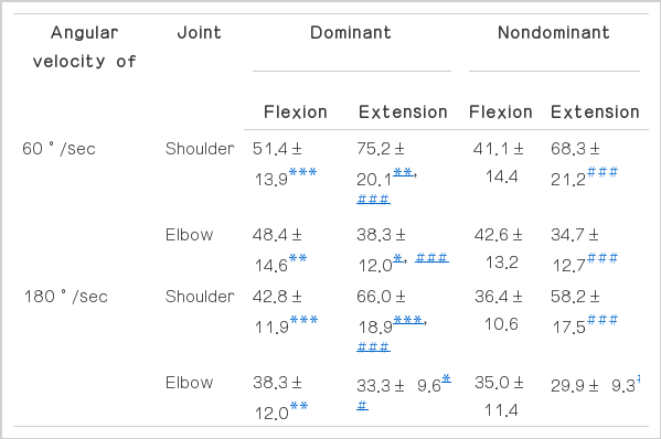
Upper limb muscular strength in wheelchair tennis players (N · m)
When measured in the condition of angular velocity of 180°/sec, shoulder joints showed higher ES score compared to FS in both arms. But elbow joints showed higher FS compared to ES and showed a tendency similar to the measured results that came from the condition of angular velocity of 60°/sec.
Also, both shoulder and elbow joints showed higher score of dominant side muscle strength compared to nondominant side muscle strength (P<0.01) (Table 2).
Ipsilateral and bilateral balance ratio
Regarding IR balance ratio (ES/FS×100) calculated with isokinetic strength measured in the condition of angular velocity of 60°/sec, shoulder joints showed the values that surpass 100% in both arms, and thus demonstrated the result of higher ES compared to FS; nondominant side showed higher score compared to dominant side (P<0.05).
Elbow joints showed less than 100% in both arms, and thus demonstrated higher FS compared to ES. There was no significant difference between both arms (Table 3). Shoulder joints showed higher IR balance ratio compared to elbow joints in both arms (P<0.001).

Upper limb ipsilateral balance ratio in wheelchair tennis players (%)
Regarding BR balance (nondominant/dominant×100) calculated with isokinetic strength measured in the condition of angular velocity of 60°/sec, shoulder joints had higher ES compared to FS (P<0.05). The same tendency was shown in elbow joints but there was no significant difference (Table 4). Also in the case of FS, elbow joints had higher BR balance compared to shoulder joints (P<0.05).
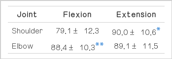
Upper limb bilateral balance ratio in wheelchair tennis players (%)
DISCUSSION
The main results of this study, regarding male wheelchair tennis players, showed that there were different tendencies of IR balance ratio between shoulder and elbow joints as demonstrated in the shoulder joints had higher ES compared to FS and that elbow joints had higher FS compared to ES. Chronic injury among wheel-chair dependents showed the highest rate in shoulder joints (Morrow et al., 2009); posterior shoulder muscle weakening and anterior shoulder muscle shortening are suggested as the sole reasons for such issue (Curtis et al., 1999). Therefore, Olenik et al. (1995) suggested exercise methods, such as rowing, backward propulsion, and exclusive retraction of the shoulder, and strengthening of rhomboideus and trapezius muscle, as prevention and treatment methods for shoulder injury.
Also, the results of this study showed significantly higher ES compared to FS in the shoulder joints of male wheelchair tennis players (P<0.001). The IR balance ratio of shoulder joint maintained the normal range (129–174%), as shown with the mean of 149–173% (Dvir, 2004; Korea Institute of Sport Science, 2003). The IR balance ratio of shoulder joint calculated from this study had a higher tendency compared to the IR balance ratio of shoulder joint of the disabled shooting players with low level of spinal cord injury are reported as 116% (Kim and Lee, 2009).
Also, regarding wheelchair basketball players, the normal range manifestation of IR balance ratio of shoulder joint [which was concluded based on the summary of studies as 187–215% of IR balance ratio of shoulder joint from Kim and Choi (2007) and Kim et al. (2002); 148–168% of IR balance ratio of shoulder joint of wheelchair tennis players from Bernard et al. (2004)] appears to be the characteristic of shoulder joint myofunction of wheelchair tennis players due to the nature of the sport.
BR balance ratio of shoulder joint for ES was 90.0% on average and thus maintained the normal range (90–110%). FS was 79.1% on average and suggested that unilateral FS strengthening exercises is necessary for nondominant shoulder joint (Davies, 1992).
Elbow joints showed significant higher FS compared to ES. Elbow joint IR balance ratio was 79.4–79.8% on average and thus did not meet the normal range (93–101%) (Dvir, 2004; Korea Institute of Sport Science, 2003). The elbow joint BR balance also had a value that did not meet the normal range (90–110%) (Davies, 1992).
Considering that traumatic injury occurrence potential increased when IR balance ratio is lower than the normal range, overuse injury occurrence potential increased when IR balance ratio is higher than the normal range, and sports injury occurrence potential due to the imbalance of muscle strength increased when BR balance is out of the normal range (Söderman et al., 2001), it can be deducted that male wheelchair tennis players have higher injury occurrence potential in elbow joints.
There is limitation to interpret the results of this study by itself due to lack of previous studies, but considering the study of Kim and Lee (2009), which reported elbow joint IR balance ratio as 110–114% and elbow joint BR balance as 90–93% among the disabled shooting players with low level of spinal cord injury, it can be deducted that the balance ratio of elbow joint muscle strength outside the normal range is caused due the characteristic of the sport of wheelchair tennis rather than caused by propulsion of wheelchair.
Regarding domestic wheelchair tennis, there is not enough infrastructure to improve the performance through scientific training, difficulty of finding players, lack of specialized trainers, lack of training systems, and limitation of facilities and equipment are suggested as the obstructions to achieve the sports performance and international competitiveness improvement (Kang, 2009; Kang and Roh, 2008). The characteristics of the expression of upper limb muscular strength of male wheelchair tennis players in this study can be used as basic data to develop sports injury prevention and exercise rehabilitation programs. It could also provide specific data on the factors of sports performance improvement.
Upon the completion of this study to understand the characteristics of the expression of upper limb muscular strength of male wheelchair tennis players in order to provide basic data for sports injury prevention and exercise rehabilitation programs development, we deducted the following conclusions.
Regarding the shoulder joints of male wheelchair tennis players, ES was significantly higher than FS (P<0.001). Also, IR balance ratio of both arms and BR balance ratio of ES were within the normal range, but BR balance ratio of FS was lower than the normal range. Therefore, the exercise intervention to strengthen FS of nondominant shoulder joint is proposed.
Regarding the elbow joints of male wheelchair tennis players, FS was significantly higher than ES (P<0.05). Also, IR balance ratio and BR balance ratio were lower than the normal range. Therefore, the exercise intervention to strengthen ES of both arms and muscle strength of nondominant elbow joint is proposed.
As a conclusion, differences between shoulder and elbow joints shown in upper limb IR balance ratio and low muscle strength balance ratio of elbow joint are considered as the characteristics of the expression of upper limb muscular strength of male wheel-chair tennis players.
Notes
CONFLICT OF INTEREST
No potential conflict of interest relevant to this article was reported.