Agreement between heart rate at first ventilatory threshold on treadmill and at 6-min walk test in coronary artery disease patients on β-blockers treatment
Article information
Abstract
The purpose of this study was to verify the accuracy of the agreement between heart rate at the first ventilatory threshold (HRVT1) and heart rate at the end of the 6-min walk test (HR6MWT) in coronary artery disease (CAD) patients on β-blockers treatment. This was a cross-sectional study with stable CAD patients, which performed a cardiopulmonary exercise test (CPET) on a treadmill and a 6-min walk test (6MWT) on nonconsecutive days. The accuracy of agreement between HRVT1 and HR6MWT was evaluated by Bland–Altman analysis and Lin’s concordance correlation coefficient (rc), mean absolute percentage error (MAPE), and standard error of estimate (SEE). Seventeen stable CAD patients on β-blockers treatment (male, 64.7%; age, 61±10 years) were included in data analysis. The Bland–Altman analysis revealed a negative bias of −0.41±6.4 bpm (95% limits of agreements, −13 to 12.2 bpm) between HRVT1 and HR6MWT. There was acceptable agreement between HRVT1 and HR6MWT (rc=0.84; 95% confidence interval, 0.63 to 0.93; study power analysis=0.79). The MAPE of the HR6MWT was 5.1% and SEE was 6.6 bpm. The ratio HRVT1/HRpeak and HR6MWT/HRpeak from CPET were not significantly different (81%±5% vs. 81%±6%, P=0.85); respectively. There was a high correlation between HRVT1 and HR6MWT (r=0.85, P<0.0001). Finally, the results of the present study demonstrate that there was an acceptable agreement between HRVT1 and HR6MWT in CAD patients on β-blockers treatment and suggest that HR6MWT may be useful to prescribe and control aerobic exercise intensity in cardiac rehabilitation programs.
INTRODUCTION
Cardiovascular diseases are the predominant cause of death worldwide and coronary artery disease (CAD) is the most common type of heart disease, accounting for 42.6% of all deaths in the United States of America (Virani et al., 2020). An essential component of contemporary care for patients with CAD is the promotion of a healthy lifestyle and control of cardiovascular risk factors (Knuuti et al., 2020). Exercise-based cardiac rehabilitation programs promote a reduction in mortality rates, decrease the risk of rehospitalization, and improve the quality of life of patients with CAD (Anderson et al., 2016; Dunlay et al., 2014). In cardiac patients, regular practice of supervised physical exercise requires an individualized prescription to optimize exercise benefits and minimize risks, offering greater safety during training (Carvalho et al., 2020; Fletcher et al., 2013).
In CAD patients undergoing cardiac rehabilitation, the recommended aerobic exercise intensity is close to the first ventilatory threshold (VT1) (Pymer et al., 2020; Tabet et al., 2008; Tan et al., 2017). The VT1 represents the exercise intensity corresponding to the transition from purely aerobic metabolism to a point where blood lactate begins to rise in the blood but reaches equilibrium (as a result of progressive activation of anaerobic glycolysis, with stable lactate levels of 1.5–2 mmol/L due to an equal lactate rate of appearance vs. rate of disappearance) (Hansen et al., 2021). Regular exercises with intensity equivalent to VT1 is medically safe (Chaloupka et al., 2005; Tabet et al., 2008), precede the ischemic threshold (Bussotti et al., 2006), elicit significant physiological adaptations (Prado et al., 2016) and clinical benefits (Zheng et al., 2008). However, the use of a cardiopulmonary exercise test (CPET), necessary to VT1 definition, involves high costs which limit its practical application in most cardiovascular rehabilitation services, mainly in low to middle-income countries. Furthermore, it is known that fewer patients are undergoing a CPET at the entry to cardiac rehabilitation (Brubaker et al., 2018; Hansen et al., 2021).
Studies have been investigating alternatives ways to prescribing aerobic exercise in heart diseases patients (Casillas et al., 2013; Casillas et al., 2017; Oliveira et al., 2016). The 6-min walk test (6MWT) is a well-established method of assessing physical capacity, practical, simple, and low-cost test, used in cardiovascular rehabilitation programs (Bellet et al., 2012; Casillas et al., 2013). Despite the well-known value of the 6MWT to predict cardiorespiratory fitness, little is known about exercise prescription intensity based on 6MWT heart rate (HR6MWT) in CAD patients on β-blockers treatment (Casillas et al., 2017).
Gayda et al. (2004) showed that VO2 and heart rate values at the end of 6MWT did not differ from those values obtained during CPET at VT1. However, in this study, some CAD patients were not on β-blockers treatment. Furthermore, the accuracy of the agreement between HR6MWT and heart rate at VT1 (HRVT1), important for use as a real clinical practice exercise control parameter, was not yet reported. Thus, the purpose of this study was to evaluate the accuracy of the agreement between HRVT1 and HR6MWT in CAD patients on β-blockers treatment. Our hypothesis is that is an acceptable agreement between heart rate from 6MWT and CPET in CAD patients on β-blockers treatment and that HR6MWT may be useful to prescribe aerobic exercise in cardiac rehabilitation programs.
MATERIALS AND METHODS
Participants
Seventeen patients with CAD under β-blockers treatment were recruited from the institutional phase II cardiovascular rehabilitation program. All participants met at least three inclusion criteria, such as acute myocardial infarction (MI), coronary artery bypass grafting (CABG), percutaneous coronary intervention (PCI), obesity, and type 2 diabetes mellitus. Exclusion criteria were diagnosis of atrial fibrillation, hemodynamic instability, ventricular arrhythmias, and respiratory, neurological, or musculoskeletal disease. In addition, patients who failed to achieve a maximum respiratory exchange ratio (>1.05) in the CPET were excluded from the analysis (Keteyian et al., 2010). All individuals provided informed written consent, and the study protocol was approved by the University of Passo Fundo institutional research ethics committee (#363/10).
Experimental procedure
Patients were undergone a CPET and after 48 hours to two 6MWTs. The CPET was performed on a treadmill (Master ATL, Inbramed, Porto Alegre, Rio Grande do Sul, Brazil) in a controlled temperature (21°C–23°C) laboratory. Individuals were continuously monitored in electrocardiographic derivations, using the ramp protocol with analysis of expired gases (O2 and CO2) in open circuits (Ergo PC Elite software, Micromed, Brasilia, Brazil) (Fletcher et al., 2013). At the beginning of each CPET day, an auto-calibration was performed according to the manufacturer’s instructions. A medium-flow pneumotachograph (10 to 120 liters/min) was used and measurements were collected in fixed time every 20 sec (VO2000, Aerosport MedGraphics, Saint Paul, MN, USA). The VT1 was determined by the ventilatory technique identifying the lower points of the oxygen ventilatory equivalent (VE/VO2) and the expired fraction of O2 before these values began to rise, while the ventilatory equivalent of carbon dioxide (VE/VCO2) and the expired CO2 fraction remained stable (Mezzani et al., 2009). The highest oxygen uptake observed in the last 40 sec of the exercise was defined as VO2peak (Fletcher et al., 2013). The heart rate at the first ventilatory threshold (HRVT1) was calculated as the average of 40 sec and heart rate response at the first ventilatory threshold (HRRVT1) was calculated as HRVT1 minus HR at rest. The age-predicted maximal HR was derived from the prediction equations HRmax=(208–0.7×age) in men and HRmax= (206–0.88×age) in women (Fletcher et al., 2013). In addition, we evaluated the HR response to incremental CPET.
The 6MWT was performed following the guidelines established by the European Respiratory Society/American Thoracic Society by trained evaluators (Holland et al., 2014). Patients walked as far as possible for six minutes in a 30-m-long flat lane, with standardized incentives. Two tests were performed with an interval of 60 min and the longest covered distance test was used for analysis (Bellet et al., 2012; Holland et al., 2014). During the 6MWT, all patients used the HR monitor (Polar S810i, Polar Electro Oy, Kempele, Finland) that continuously showed the HR throughout the 6-min test. The highest value of the HR was noted during the last 30 sec of the 6MWT (HR6MWT) (Morard et al., 2015). Resting HR was assessed while the patients were standing for 60 sec, after a 5-min rest sitting. The HR6MWT was used to assess the relative intensity of the 6MWT to the maximal HR from CEPT. Heart rate response at the end of the 6MWT (HRR6MWT) was calculated as HR6MWT minus HR at rest. The predicted distance was calculated using the formula (equation 1) proposed by (Britto et al., 2013). All tests were performed between 8:00 and 11:00 a.m., at the same place, and followed the same protocol.
Statistical analysis
Data distribution was evaluated by the Shapiro–Wilk test and expressed in mean, standard deviation (SD), and qualitative variables by frequency distribution. Comparisons between HRVT1 and HR6MWT were performed by paired t-test. Pearson correlation and Bland–Altman analyses were used to evaluate the association and agreement between HRVT1 and HR6MWT, respectively (Bland and Altman, 1986). Limits of agreement were calculated (mean bias± SD×1.96) for each Bland–Altman assessment. Mean absolute percentage error (MAPE) [(HRVT1–HR6MWT)/HRVT1×100] provided a general measurement error for HR6MWT. In addition, the accuracy of agreement between HRVT1 and HR6MWT was evaluated by Lin concordance correlation coefficient (rc) (Lin, 1989) and by the standard error of estimate (SEE) (Sarzynski et al., 2013). We considered an acceptable accuracy of agreement when rc>0.8 (Gillinov et al., 2017), MAPE within 5% (Navalta et al., 2020) and SEE ≤7 bpm (Oliveira et al., 2016). P values <0.05 were considered significant. Data were analyzed using GraphPad Prism 6 (GraphPad Software, La Jolla, CA, USA) and IBM SPSS Statistics ver. 19.0 (IBM Co., Armonk, NY, USA). Lin concordance correlation coefficient study power analysis was calculated using Power Analysis & Sample Size 2021 (NCSS, LLC, Kaysville, UT, USA). This statistical test uses a one-sided z-test with a 0.15 significance level.
RESULTS
Seventeen patients (64.7% men), 61±10 years old, and 29± 5 kg/m2 of body mass index were included in the study. Most patients had a diagnosis of MI with primary (53%) or elective (23.5%) PCI, CABG (23.5%), and all patients were under optimal medical treatment. Twelve patients used selective β1-adrenergic receptor antagonists (54±23 mg/day); four patients used the third generation of β-blockers with vasodilatory effect (15±6 mg/day) and one patient used propranolol (160 mg/day). Additional patients’ clinical characteristics are shown in Table 1.
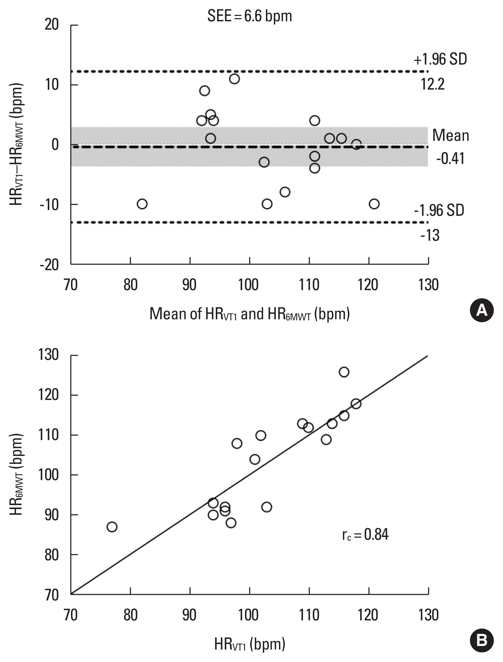
Table 2 shows the CPET and the 6MWT results. All patients performed both tests without complications. No statistically significant differences were found between HRVT1 and HR6MWT (103.2± 10.8 bpm vs. 103.6±12.3 bpm, P=0.81). When expressed in percentage of CPET HRmax, HRVT1/HRpeak and HR6MWT/HRpeak were not statistically different (81%±5% vs. 81%±6%, P=0.85). On average, HR6MWT’s MAPE was 5.1%, and SEE was 6.6 bpm. Six patients (35%) showed a difference higher than five bpm between HRVT1 and HR6MWT.
Bland–Altman analysis of agreement between HRVT1 and HR6MWT revealed a negative bias of −0.41±6.4 bpm and 95% limits of agreements, −13 to 12.2 bpm (Fig. 1A). Pearson correlation showed a high and significant association between HRVT1 and HR6MWT (r=0.85, P<0.0001). In addition, Lin concordance correlation coefficient provided acceptable agreement (rc=0.84; 95% CI, 0.63 to 0.93; study power analysis=0.79) (Fig. 1B). These results suggest that HR6MWT reflects HRVT1 accurately in CAD patients using β-blockers.

Assessment of the agreement between the HRVT1 and the HR6MWT for patients with coronary artery disease. (A) Bland–Altman plot shows the difference between HRVT1 and the HR6MWT (y-axis). The middle line represents the mean difference between methods and shaded areas present confidence interval limits for the mean (95% confidence interval [CI], −3.7 to 2.9 bpm). The 95% limits of agreement (±1.96 standard deviation [SD]) fall within −13 to 12.2 bpm. (B) Concordance correlation coefficient shows agreement of HRVT1 with HR6MWT (95% CI, 0.63 to 0.93). HR6MWT, heart rate at 6th min of 6-min walk test; HRVT1, heart rate at the first ventilatory threshold; SEE, standard error of estimate; rc, Lin concordance correlation coefficient.
DISCUSSION
The main result of the present study demonstrates an acceptable agreement between HRVT1 and HR6MWT and, combined with smaller values of the MAPE and SEE, point out that is possible to use HR6MWT as an equivalent as HRVT1 to prescribe and monitor the aerobic exercise in CAD patients on β-blockers treatment.
Bland–Altman analysis results indicate the agreement between HRVT1 and HR6MWT values. Furthermore, the addition of SEE and MAPE to Bland–Altman analysis permitted us to measure the accuracy of agreement between HRVT1 and HR6MWT. Thus, our results showed that included patients meet the criterion of the acceptable concordance between HRVT1 and HR6MWT (SEE≤7 bpm and MAPE≤5%) (Navalta et al., 2020; Oliveira et al., 2016). Obtained data intervals are within an acceptable range for training with HR-derived methods (≤8 bpm) (Robergs and Landwehr, 2002). Our results agree and reinforce existent literature. Morard et al. (2015) reported agreement between HRVT1 in a symptom-limited incremental CPET on a cycle ergometer and HR6MWT in CAD patients with an acceptable 95% confidence interval of bias from Bland–Altman analysis (−12.3 to 8.1 bpm). Similarly, in patients with chronic heart failure, Oliveira et al. (2016) showed a high correlation between HRVT1 and HR6MWT (r=0.81; P<0.0001), with an acceptable SEE of 6.05 bpm. In clinical practice, our results propose the HR6MWT may be used to individualize and improve aerobic exercise prescriptions particularly to CAD patients under β-blockers treatment. Thus, it is expected that the prescription of more accurate aerobic exercises may enhance the beneficial effects of aerobic training in patients undergoing a cardiac rehabilitation program.
Previous data suggest that adding ~30 bpm to resting HR is a simple way to infer HRVT1 values when a CPET was not performed (Nemoto et al., 2019). Although our HRRVT1 values (31±11 bpm) were close to the suggested values, we believe that performing the 6MWT may be a more accurate form of exercise prescription. In addition, the HRR6MWT was 30±8 bpm and represents 55% of HRRpeak, a moderate aerobic exercise intensity (Hansen et al., 2019; Pelliccia et al., 2021).
The HRVT1 and HR6MWT were close to 80% of the CPET HRpeak. This percentage may be considered as moderate to high intensity and is in line with the recommended aerobic training zone (~75% to ~88% HRpeak) reported in two large cohorts of patients with CVD (Díaz-Buschmann et al., 2014; Hansen et al., 2019). Furthermore, Díaz-Buschmann et al. (2014) recommend that patients on β-blockers treatment exercise at 80% HRpeak, reinforcing the clinical applicability of our results. Additionally, clinical safety must be considered in exercise prescription in CAD patients. Thus, the founded intensity, ~80% CPET HRpeak, agree with recommended safety limit for aerobic exercise (<90% HRpeak) (Pelliccia et al., 2021).
Although Lin concordance correlation coefficient calculated study power provided acceptable agreement, is possible that this study has limitations inherent to the sample size. Furthermore, as we did not have an equal distribution between the different β-blockers used in CAD treatment the interpretation of the present results must be performed with prudence and may restrict the extrapolation of the results to another evaluated sample. Finally, as both CPET and 6MWT were performed with patients walking, our results are more accurate to this kind of aerobic exercise than the cycle ergometer, or another exercise to lower limbs aerobic exercises.
Our results suggest that the highest heart rate observed during the last 30 sec of the 6MWT seems to be a valid tool to prescribe aerobic exercise intensity with safe at entry in a cardiac rehabilitation program for β-blockers patients. More research should be carried out to evaluate the use of HR6MWT to control the exercise intensity in the progression of the physical training program in cardiac rehabilitation.
ACKNOWLEDGMENTS
The authors received no financial support for this article.
Notes
CONFLICT OF INTEREST
No potential conflict of interest to this article was reported.


